Bio Science
- Home / Bio Science
- MatTek Human 3D Tissue (인공피부)

MatTek’s advanced 3D full thickness model of Human Skin takes preclinical testing to the next level by providing a physiological means to evaluate early molecular markers of efficacy.
제품 상세정보
The development of innovative skincare products requires advanced, human-relevant in vitro testing technologies. When world-leading personal care and cosmetic scientists evaluate novel ingredients and formulations, they choose the EpiDermFT 3D human skin model.


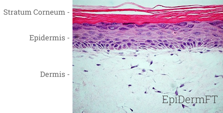
Engineered to enable in vitro skin research in which fibroblast-keratinocyte cell interactions are important, MatTek’s EpiDermFT system consists of normal, human epidermal keratinocytes (NHEK) and normal, human dermal fibroblasts (NHFB) cultured to form a multilayered model of the human dermis and epidermis. Cultured at the air-liquid interface in easy-to-handle tissue culture inserts, EpiDermFT attains levels of differentiation at the cutting edge of tissue engineering technology.
EpiDermFT consists of organized keratin 5 expressing basal cells, involucrin and keratin 10 expressing spinous and granular layers, and cornified epidermal layers analogous to those found in vivo. The dermal compartment is composed of a collagen matrix containing viable normal human dermal fibroblasts (NHDF). The epidermal and dermal layers are mitotically and metabolically active and exhibit in vivo-like morphological and growth characteristics which are uniform and highly reproducible.

A well-developed basement membrane is present at the dermal/epidermal junction. Hemidesomosomes(›), lamina lucida, lamina densa(→) and anchoring fibril structures(») are evident by transmission electron microscopy. |
|

Kit: A standard EpiDermFT kit (EFT-400) consists of 24 tissues. (Tissue “kits” contain tissues, a small amount of culture medium, and plasticware; contact MatTek for specific kit contents)
Substrate: Costar Snapwell™ single well tissue culture plate inserts are used. Pore size = 0.4 μm, Diameter = 1.2 cm. Surface area = 1.0 cm2
Culture: Air-liquid interface
Histology: 8-12 cell layers plus stratum corneum (basal, spinous, and granular layers)
Lot numbers: Tissue lots produced each week are assigned a specific lot number. A letter of the alphabet is appended to the end of the lot number to differentiate between individual kits within a given lot of tissues. All tissue kits within a lot are identical in regards to cells, medium, handling, culture conditions, etc.
Shipment: At 4°C on medium-supplemented, agarose gels in 6-well plates
Shipment day: Every Monday. Shipment on Thursday also possible upon special request
Delivery: Tuesday morning via FedEx priority service (US). Outside US: Tuesday-Thursday depending on location
Shelf life: Including time in transit, tissues may be stored at 4°C for up to 6 days prior to use. However, extended storage periods are not recommended unless necessary. In addition, the best reproducibility will be obtained if tissues are used consistently on the same day, e.g. Tuesday afternoon or following overnight storage at 4°C (Wednesday morning)
Length of experiments: Cultures can be continued for up to 2 weeks with good retention of normal epidermal morphology. Cultures must be fed every other day with 5.0 ml of EFT-400 medium
Type: Normal human epidermal keratinocytes (NHEK); Normal human dermal fibroblasts (NHDF)
Genetic make-up: Single donor
Derived from: Neonatal-foreskin tissue (NHEK); Neonatal skin (NHDF)
Alternatives: NHEK from adult breast skin; NHDF from adult skin
Screened for: HIV, Hepatitis-B, Hepatitis-C, mycoplasma
Base medium: Dulbecco’s Modified Eagle’s Medium (DMEM)
Growth factors/hormones: Epidermal growth factor, insulin, hydrocortisone and other proprietary stimulators of epidermal differentiation
Serum: None
Antibiotics: Gentamicin 5 µg/ml (10% of normal gentamicin level)
Anti-fungal agent: Amphotericin B 0.25 µg/ml
pH Indicator: Phenol red
Other additives: Lipid precursors used to enhance epidermal barrier formation (proprietary)
Alternatives: Phenol red-free (EFT-400-PRF), antibiotic-free (EFT-400-ABF), anti-fungal-free (EFT-400-AFF), or hydrocortisone-free medium and tissue (EFT-400-HCF) are available. Agents are removed at least 3 days prior to shipment
Assay/Maintenance medium: EFT-400-ASY is utilized for assays; EFT-400-MM is used for term maintenance of the EFT-400 tissues
Visual inspection: All tissues are visually inspected and if physical imperfections are noted, tissues are rejected for shipment
End-use testing: Tissues are exposed to 1% Triton X-100 for 8, 12, 16 and 24 hours. The time of exposure required to reduce the tissue viability (ET-50) using the MTT viability assay is determined (See MatTek EpiDermFT use protocol) for each lot of tissue. ET-50’s generally fall within the range of 6.5-9.5 hours. ET-50’s in customers’ lab may differ slightly from the MatTek results
Sterility: All media used throughout the production process is checked for sterility. Maintenance medium is incubated with and without antibiotics for 1 week and checked for sterility. The agarose gel from the 24-well plate used for shipping is also incubated for 1 week and checked for any sign of contamination
Screening for pathogens: All cells are screened and are negative for HIV, hepatitis B and hepatitis C using PCR. However, no known test method can offer complete assurance that the cells are pathogen free. Thus, these products and all human derived products should be handled at BSL-2 levels (biosafety level 2) or higher as recommended in the CDC-NIH manual, “Biosafety in microbiological and biomedical laboratories,” 1998. For further assistance, please contact your site Safety Officer or MatTek technical service
Notification of lot failure: If a tissue lot fails our QC or sterility testing, the customer will be notified and the tissues will be replaced without charge when appropriate. Because our QC and sterility testing is done post-shipment, notification will be made as soon as possible (Under normal circumstances, ET-50 failures will be notified by Wednesday 5 p.m.; sterility failures will be notified within 8 days of shipment)

EpiDermFT tissues are used across a wide range of applications including scientific efficacy and claims substantiation. Due to a dry skin surface, EpiDermFT can be used to evaluate biological responses to topical applications of formulations or ingredients. Simple protocols and the evaluation of early molecular endpoints allow research to acquire data in days, not weeks or months.
Examine changes in extracellular matrix gene and protein expression and histological structure to determine the anti-aging efficacy of cosmetic ingredients or final formulation. EpiDermFT is available from a range of adult donors.
Use EpiDermFT to evaluate therapeutics with the wound healing assay kit. Wound healing assay kits include 24 pre-wounded tissues and are available with epidermal only wounds or full-thickness wounds of the epidermis and dermis.
Use EpiDermFT to assess the efficacy of topically applied moisturizers by measuring the electrical impedance of the epidermis. Amenable to creams, gel, lotions or other formulations, the Skin Hydration assay allows for data analysis within 24 hours.
Skin Hydration Application Note
Analyze cyclopyrimidine dimer formation, inflammatory mediator release and oxidative endpoints to evaluate the UV protective efficacy of compounds or formulations.
UV Protection Application Note
Browse our reference library to see how our EpiDermFT tissue has been used in these areas of study.
4 Neonatal & 6 Adult w/ Matched NHEK and NHDF